

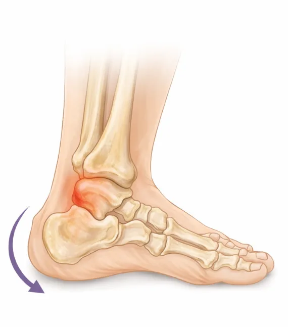
Correction of foot alignment in hindfoot deformities
Hindfoot deformities involve malalignment of the calcaneus and talus and directly affect the axis of the entire lower limb. When the heel shifts inward or outward, weight-bearing balance is disrupted, ankle instability may develop, and stress on ligaments and tendons increases. Over time, this can lead to chronic pain, fatigue while walking, and reduced physical activity.
The hindfoot plays a key role in establishing a stable gait and maintaining balance. It determines the direction of foot progression during weight-bearing, and any axis deviation influences overall walking mechanics. In pronounced deformities, the body compensates by altering the function of the knee and hip joints, increasing the risk of secondary problems.
Surgical correction of hindfoot deformities is aimed at restoring proper alignment and stable support. We individualize the treatment strategy based on the type of deformity, joint condition, and ligament integrity. This approach addresses the underlying cause of instability, reduces painful load, and creates conditions for confident and safe walking without progression of the deformity.
How the hindfoot affects gait and body balance
The hindfoot plays a crucial role in maintaining balance and establishing proper gait mechanics. The position of the calcaneus determines the direction of weight-bearing with each step and influences load distribution throughout the lower limb. When hindfoot alignment is disrupted, movements become less stable and body balance is more difficult to control.
Changes in heel position affect not only the ankle joint but also the biomechanics of the knee and hip. The body compensates for axis deviations by altering gait patterns, which over time can lead to overload of joints and muscles higher in the kinetic chain. These changes are often accompanied by a sense of instability while walking and increased fatigue.
We assess how the hindfoot contributes to balance and load transfer during movement. Gait analysis and evaluation of bone alignment help determine the extent of axis deviation and its impact on the entire lower limb. Correcting hindfoot alignment helps restore a stable gait, improve movement control, and reduce stress on the joints involved in walking.
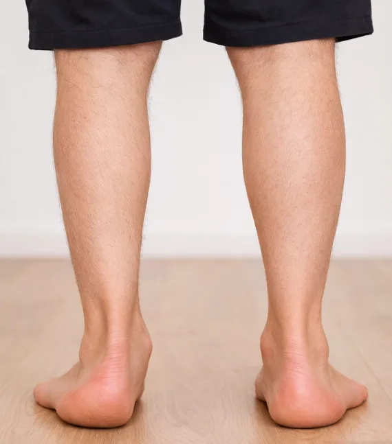
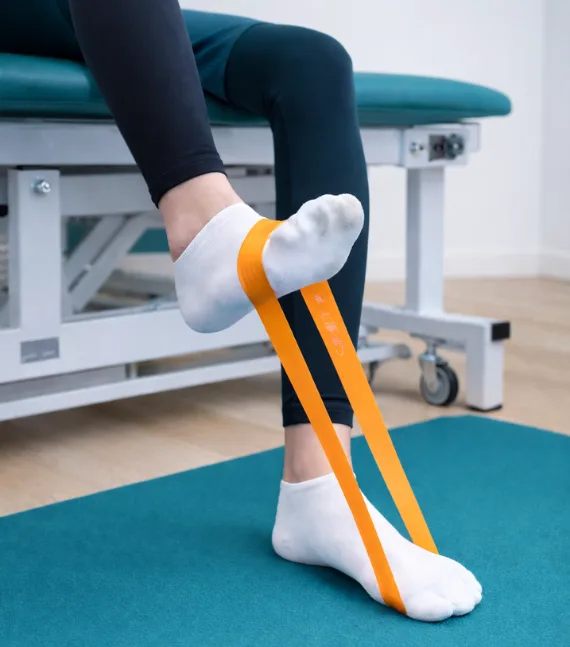
Restoration of weight-bearing and stability after hindfoot correction
After surgical correction of hindfoot deformities, the primary goal of treatment is to restore stable weight-bearing and confidence while walking. Proper positioning of the calcaneus and realignment of the foot axis reduce overload on joints and ligaments that previously experienced excessive strain. This creates the foundation for a more stable and predictable gait.
During recovery, it is essential to gradually adapt the foot to load and establish correct movement patterns. Controlled progression of activity helps prevent recurrent displacement and strengthens the muscles responsible for foot stabilization. This approach is particularly important for patients who previously experienced instability or pain during prolonged walking.
We support the patient throughout all stages of rehabilitation, assessing foot stability, load distribution, and comfort during movement. Structured and consistent rehabilitation helps restore balance, reduce the risk of recurrence, and preserve the long-term functional outcome of hindfoot correction.
Stages of treatment
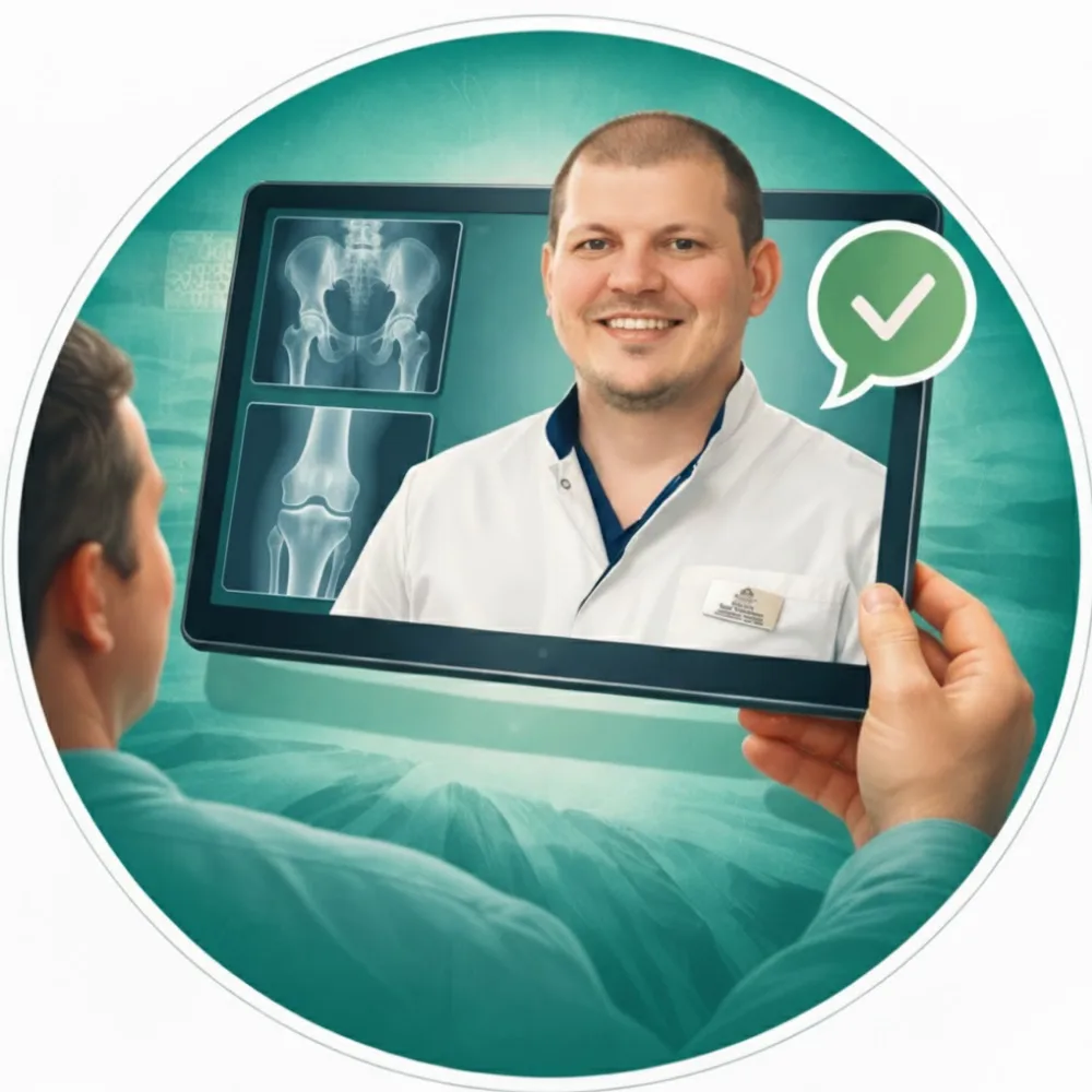
Online consultation
An online consultation allows you to discuss your condition, complaints, and symptoms in advance. You can submit imaging studies and medical documents, ask questions, and receive a preliminary treatment plan before visiting the clinic.
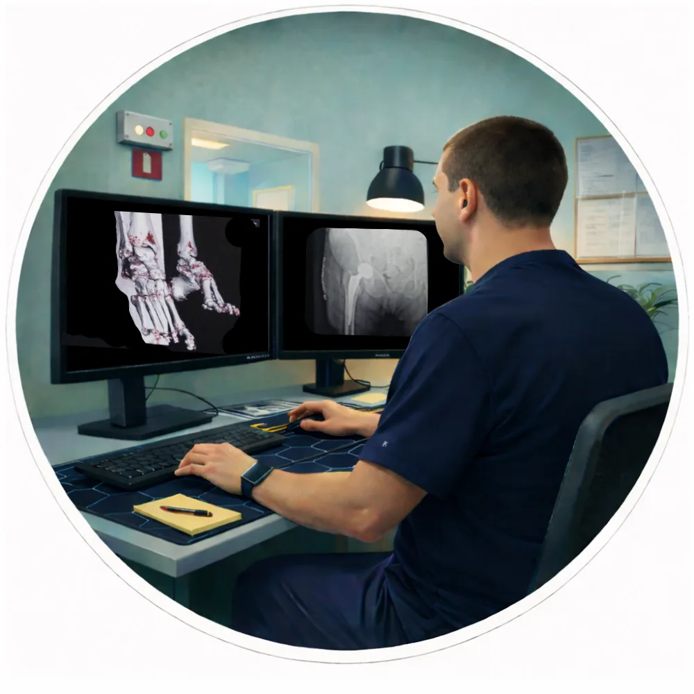
Examination and 3D surgical planning
All necessary examinations are performed within one day. A CT scan of the joint is conducted, followed by computer-based 3D surgical planning and implant selection to ensure maximum precision.

Surgery
On the second day of the clinic stay, the surgical procedure is performed. Original implants by Stryker Corporation (USA) and the MAKO robotic-assisted system are used, ensuring a high level of precision in implant positioning.
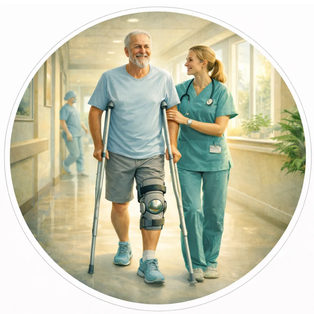
Rehabilitation and follow-up
Recovery begins immediately after surgery. In most cases, you will be able to start walking on the day of the procedure. After discharge, we continue to provide online follow-up care, including recovery guidance, rehabilitation monitoring, and answers to any questions that may arise.
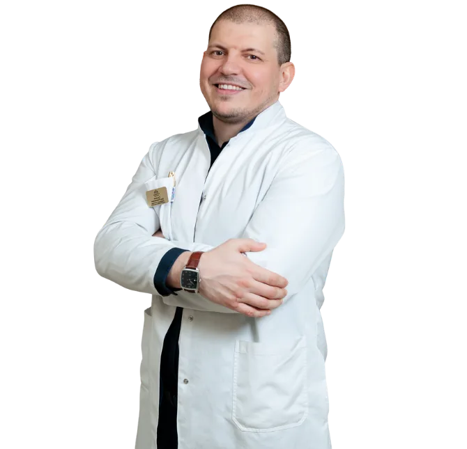
The surgery is performed personally by Zaur Sulumov
The doctor is personally involved in every stage of treatment — from the initial consultation and surgical planning to recovery monitoring and outcome evaluation.
Start Treatment
FAQ
- Calcaneal deformity
Post-fracture conditions
Severe pes planovalgus deformity
Subtalar joint osteoarthritis
Achillobursitis, contractures
- Corrective calcaneal osteotomies
Subtalar joint arthrodesis
Tendon reconstruction
Achilles tendon lengthening
Most commonly:
Cast — 4–6 weeks
No weight-bearing
After that — transition to a brace for another 2–4 weeks.
Usually after 8–10 weeks — once bone healing has been confirmed.
Full functional recovery may take 4–6 months.