

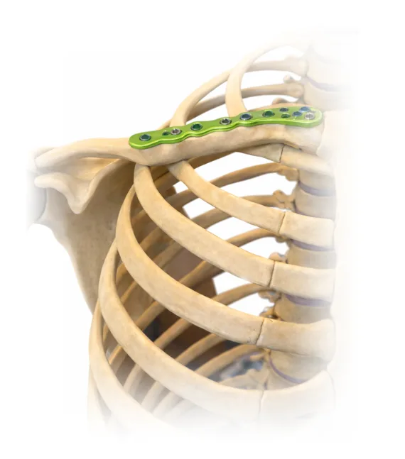
Osteosynthesis for clavicle fracture
Osteosynthesis for clavicle fracture is indicated when conservative treatment does not allow reliable restoration of bone anatomy and function. The clavicle plays a key role in shoulder girdle mechanics, providing stability and transmitting load from the upper limb to the trunk. When injured, arm biomechanics are disrupted, and improper healing may lead not only to visible deformity but also to persistent limitation of motion, decreased strength, rapid fatigue, and the development of chronic pain.
We treat patients with displaced clavicle fractures, bone shortening, instability at the fracture site, and cases where early functional recovery of the upper limb and preservation of shoulder symmetry are essential. This approach is particularly important for active individuals, athletes, and working-age patients who aim to return to their previous level of activity without pain or functional limitations.
During osteosynthesis, precise alignment of bone fragments is performed, followed by stable fixation while maintaining the correct clavicular length and axis. This creates optimal conditions for predictable bone healing and reduces the risk of secondary deformity and overload of the shoulder joint. The decision to proceed with surgical treatment is made individually after assessing the fracture pattern, imaging findings, clinical presentation, and the patient’s functional needs. At every stage of treatment, we provide guidance, explain the chosen strategy, and monitor recovery until full return to activity.
Features of clavicle fractures influencing treatment strategy
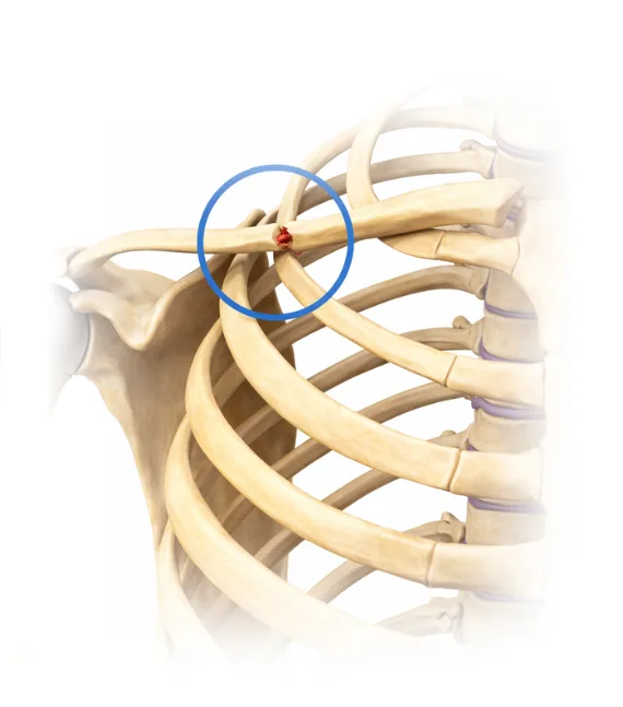
Clavicle fractures have several anatomical and biomechanical features that directly influence the choice of treatment strategy and the prognosis for recovery. The clavicle connects the upper limb to the chest wall and plays a key role in maintaining proper shoulder girdle alignment. In fractures of the middle third of the bone, displacement of fragments often occurs due to muscle forces and the weight of the arm, leading to clavicular shortening and imbalance of the shoulder girdle. Even relatively minor displacement can, over time, result in discomfort, shoulder asymmetry, and overload of the shoulder joint.
We carefully assess the fracture pattern, stability of bone fragments, and the impact of the injury on shoulder function and the entire upper limb. This approach allows us to select the most appropriate treatment strategy for each specific clavicle fracture rather than applying universal solutions, thereby reducing the risk of malunion, chronic pain, and persistent functional limitations. We also evaluate how the fracture affects the patient’s daily activities and the functional demands of recovery. This enables us to plan treatment in advance, taking into account lifestyle and expected physical нагрузку. As a result, the chosen strategy focuses not only on bone healing but also on preserving full arm function.
Цель лечения и восстановление функции при переломе ключицы
The goal of treating a clavicle fracture is not only to achieve bone union, but also to restore full function of the shoulder girdle and upper limb. Even when radiographic healing is achieved, improper clavicular alignment may lead to restricted range of motion, reduced arm strength, and overload of the shoulder joint. Therefore, in managing clavicle fractures, it is essential to consider not only imaging findings but also future biomechanics, shoulder symmetry, and the patient’s comfort in daily life.
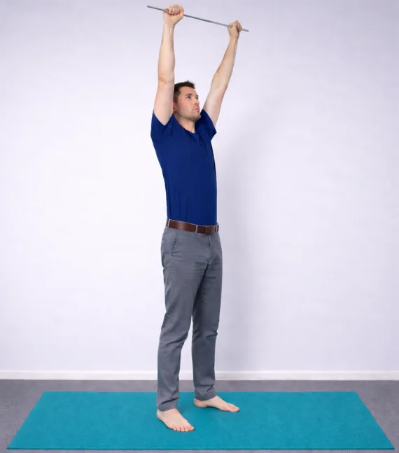
After clavicle osteosynthesis, careful monitoring of recovery and gradual return to load are crucial. We focus on restoring shoulder range of motion, coordination, and arm endurance, as well as reducing the risk of chronic pain and functional limitations. This strategy helps prevent late complications and achieve a stable long-term outcome.
We support the patient throughout the recovery phase, assessing bone healing progress and adjusting recommendations according to individual goals and activity level. This approach allows treatment to be structured in a way that enables the patient to return to normal life without stiffness, pain, or movement restrictions in the shoulder girdle.
Stages of treatment
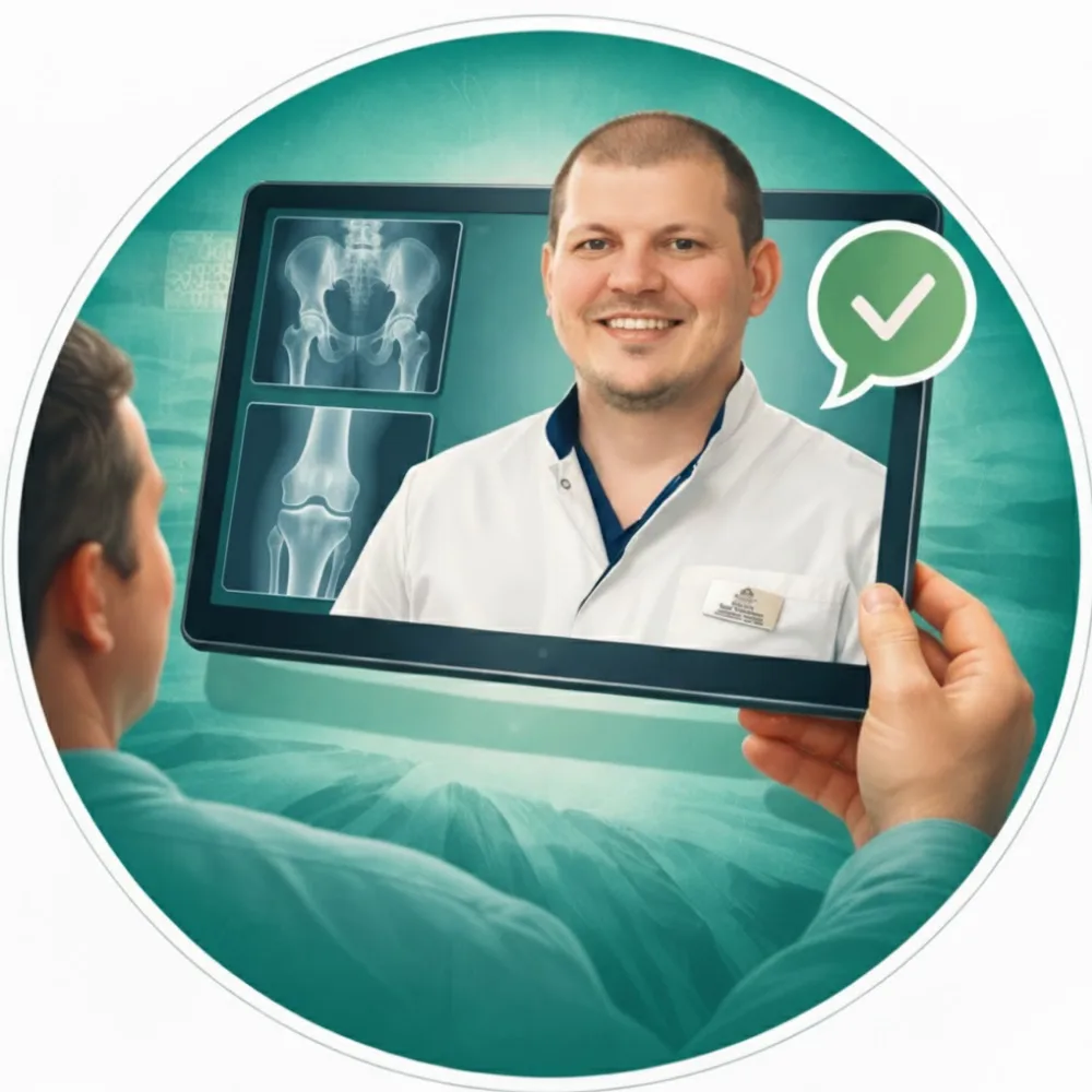
Online consultation
An online consultation allows you to discuss your condition, complaints, and symptoms in advance. You can submit imaging studies and medical documents, ask questions, and receive a preliminary treatment plan before visiting the clinic.
Examination and 3D surgical planning
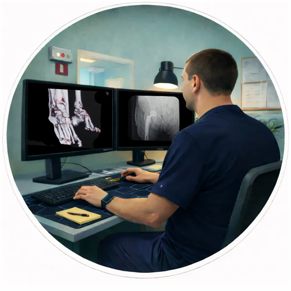
All necessary examinations are performed within one day. A CT scan of the joint is conducted, followed by computer-based 3D surgical planning and implant selection to ensure maximum precision.

Surgery
On the second day of the clinic stay, the surgical procedure is performed. Original implants by Stryker Corporation (USA) and the MAKO robotic-assisted system are used, ensuring a high level of precision in implant positioning.

Rehabilitation and follow-up
Recovery begins immediately after surgery. In most cases, you will be able to start walking on the day of the procedure. After discharge, we continue to provide online follow-up care, including recovery guidance, rehabilitation monitoring, and answers to any questions that may arise.
The surgery is performed personally by Zaur Sulumov
The doctor is personally involved in every stage of treatment — from the initial consultation and surgical planning to recovery monitoring and outcome evaluation.
Start Treatment
FAQ
The duration depends on the type of fracture and the fixation method. On average, surgery lasts from 30 to 120 minutes. In complex or comminuted fractures, operative time may be longer.
The timing of weight-bearing depends on the fracture location and stability of fixation. In some cases, movement is allowed within the first days after surgery, while full weight-bearing depends on the fracture type. For intra-articular fractures, a forced pause of 4–8 weeks may be required, with gradual progression under medical supervision and follow-up X-rays.
There is no pain during the operation due to anesthesia. After surgery, discomfort is managed with a modern pain-control protocol and typically decreases as the fracture heals.
Not always. Implants from leading manufacturers are commonly used, and in most cases plates and screws can remain in the body permanently. Removal is considered individually if there is discomfort or specific medical indications.
Average bone healing time ranges from 6 to 12 weeks, but this may vary depending on the patient’s age, fracture type, and overall health condition.
Rehabilitation focuses on restoring movement, muscle strength, and limb function. The program is individualized and may include physical therapy, physiotherapy, and gradual increase of load.
Not all fractures require surgical treatment. However, in cases of fragment displacement, instability, or risk of improper healing, osteosynthesis ensures accurate alignment and improves the functional outcome.