Recovery takes 3–4 months, sometimes longer — in the case of arthrodeses.


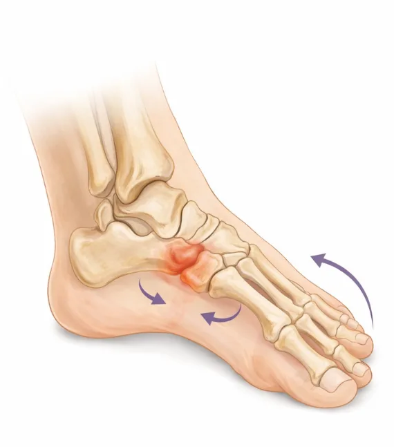
Surgical correction of midfoot deformities
Deformities of the midfoot often develop gradually and may remain underestimated for a long time. Pain in the arch region, a sense of instability while walking, rapid fatigue, and reduced endurance are early signs of impaired weight-bearing function. Over time, these changes can lead to flatfoot deformity, osteoarthritis of the midfoot joints, and chronic pain.
The midfoot plays a key role in maintaining the longitudinal and transverse arches and in transferring load between the hindfoot and forefoot. When deformity occurs, the coordinated function of the entire foot is disrupted, resulting in uneven load distribution. This increases stress on the joints and ligaments, reduces shock absorption, and makes gait less stable.
Surgical correction of midfoot deformities is aimed at restoring anatomical alignment and stable support. We individualize the treatment strategy based on the cause of deformity, joint condition, and the degree of functional impairment. This approach not only reduces pain but also creates conditions for long-term restoration of weight-bearing function without progression of the deformity.
The role of the midfoot in arch support and shock absorption
The midfoot serves as a connecting link between the heel and the forefoot, providing arch stability and shock absorption with each step. This region bears a significant portion of the load during the stance phase, when body weight shifts forward. When the structure of the midfoot is compromised, the foot loses its ability to effectively absorb impact, which affects walking comfort and endurance.
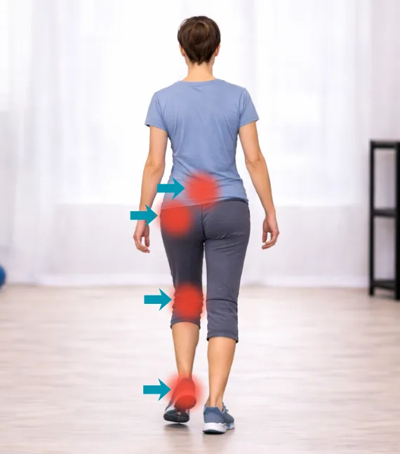
Deformities and osteoarthritis of the midfoot joints reduce mobility and alter arch function. As a result, the foot becomes more rigid, adapts less effectively to uneven surfaces, and fatigues more quickly during prolonged activity. These changes are often accompanied by pain not only in the foot but also in the knees and hips due to compensatory gait alterations.
We assess how the midfoot contributes to load distribution and shock absorption during movement. Biomechanical analysis helps identify overloaded joints and ligaments that require correction. Restoring proper midfoot function improves gait stability, reduces stress on proximal joints, and enhances overall movement quality.
Restoration of stability and weight-bearing function of the foot
After surgical correction of midfoot deformities, the key objective is to restore stable support and the foot’s ability to bear weight without pain. Proper alignment of the bones and joints helps reestablish coordinated arch function and reduce excessive strain on the soft tissues that developed as a result of the deformity.
The recovery phase focuses on gradual adaptation of the foot to load and the formation of a stable gait pattern. As tissues heal, it is important not only to restore mobility where necessary but also to ensure stability in weight-bearing zones. This balance helps prevent overload of specific areas of the foot and reduces the risk of recurrent pain during walking.
We support the patient throughout rehabilitation, assessing foot stability, load distribution, and comfort during movement. A structured and consistent approach to recovery helps restore confidence while walking, improve endurance, and preserve the long-term results of surgical correction.
Stages of treatment
01
Online consultation
An online consultation allows you to discuss your condition, complaints, and symptoms in advance. You can submit imaging studies and medical documents, ask questions, and receive a preliminary treatment plan before visiting the clinic.
02
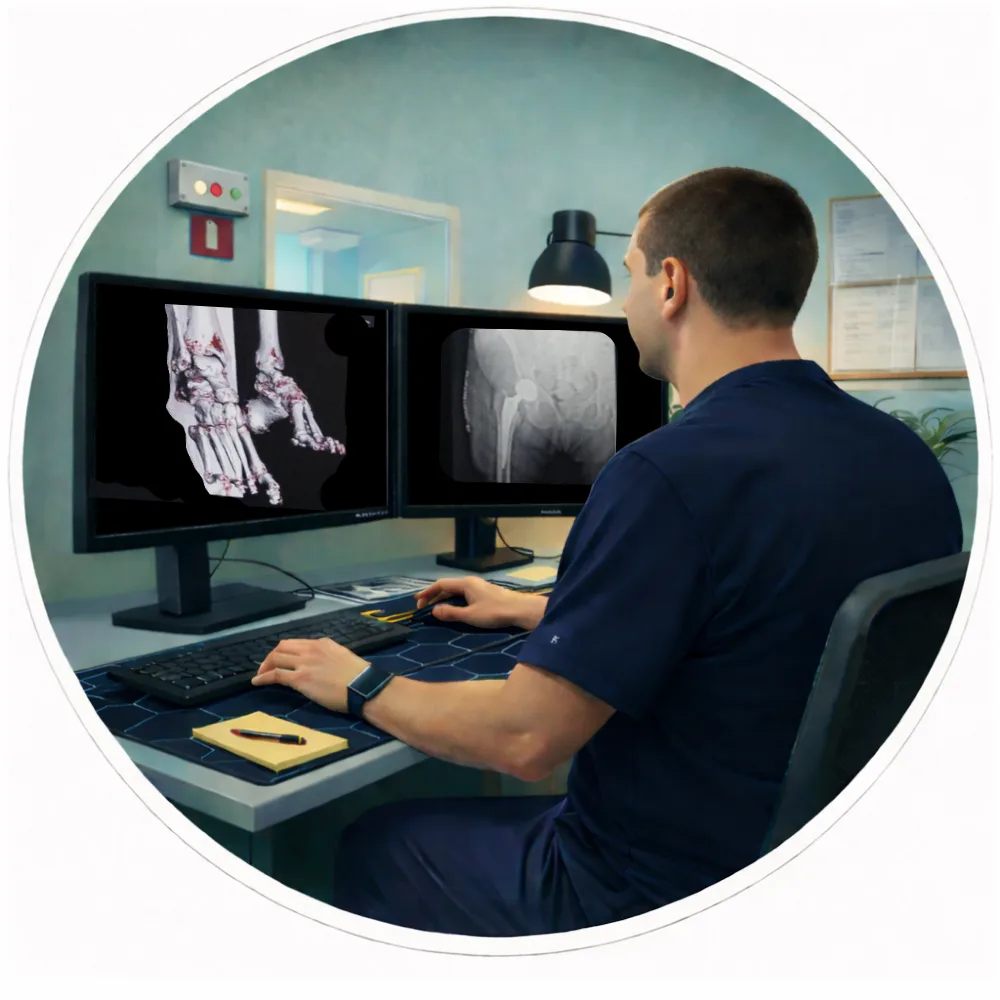
Examination and 3D surgical planning
All necessary examinations are performed within one day. A CT scan of the joint is conducted, followed by computer-based 3D surgical planning and implant selection to ensure maximum precision.

03
Surgery
On the second day of the clinic stay, the surgical procedure is performed. Original implants by Stryker Corporation (USA) and the MAKO robotic-assisted system are used, ensuring a high level of precision in implant positioning.
04
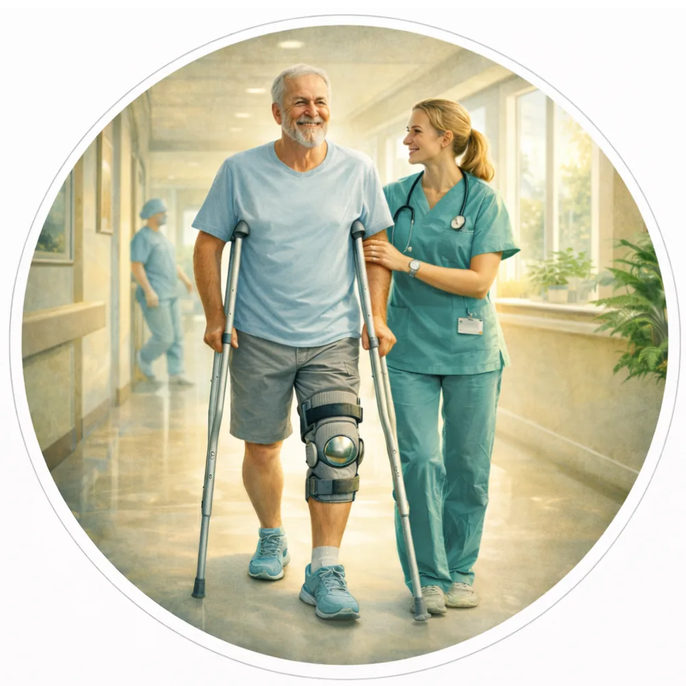
Rehabilitation and follow-up
Recovery begins immediately after surgery. In most cases, you will be able to start walking on the day of the procedure. After discharge, we continue to provide online follow-up care, including recovery guidance, rehabilitation monitoring, and answers to any questions that may arise.
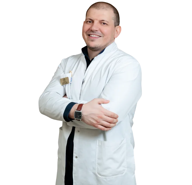
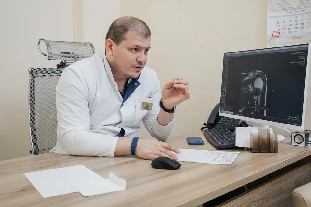
The surgery is performed personally by Zaur Sulumov
The doctor is personally involved in every stage of treatment — from the initial consultation and surgical planning to recovery monitoring and outcome evaluation.
Start Treatment
FAQ
Osteoarthritis of the tarsal joints
Pes planovalgus deformity
Post-traumatic conditions
Ligament instability
Arthrodesis (joint stabilization)
Osteotomies
Ligament reconstruction
Longitudinal arch correction
The choice depends on the cause of pain and the degree of deformity.
Most often:
Cast or rigid immobilization — 4–6 weeks
No full weight-bearing
Then transition to an orthosis or weight-bearing depending on the situation.
Partial weight-bearing — after 4–6 weeks.
Full weight-bearing — after consolidation is confirmed on X-ray (usually 8–10 weeks).
Surgery costs from $3,170