

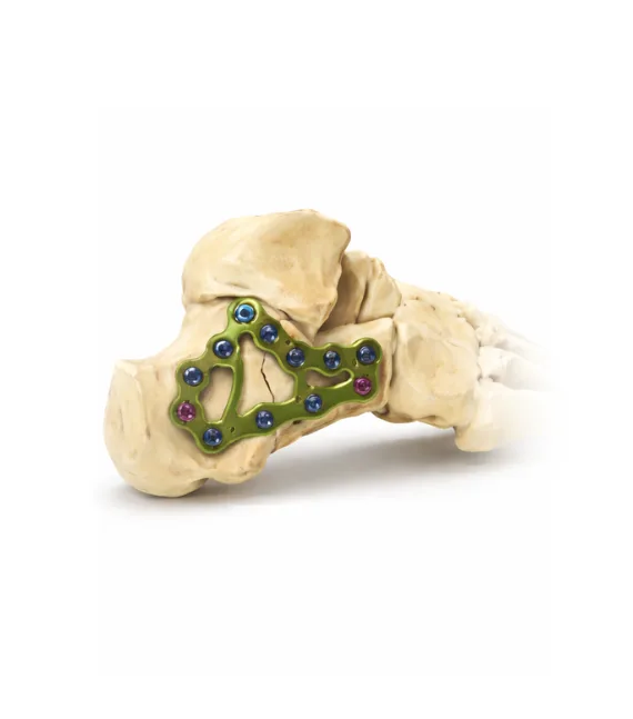
Osteosynthesis for a calcaneal fracture
Osteosynthesis for a calcaneal fracture is indicated when the injury disrupts the shape of the heel and the normal distribution of load across the foot. The calcaneus is the primary weight-bearing structure during walking, and its damage is often associated with changes in heel height and width, affecting gait and weight transfer. In displaced calcaneal fractures, especially those involving the subtalar joint or deforming the articular surface, the risk of chronic pain and persistent movement limitations significantly increases.
We treat patients with calcaneal fractures when precise restoration of the bone’s anatomical shape and preservation of hindfoot function are essential. During osteosynthesis, the goal is not only to fix the bone fragments but also to restore correct joint alignment, which is critical for shock absorption and stability during walking. This approach creates conditions for reliable healing and reduces the risk of prolonged pain under load.
The choice of surgical strategy is made individually after evaluating the fracture pattern, imaging findings, and clinical presentation. We support patients throughout all stages of treatment, monitoring recovery and helping restore weight-bearing capacity and confident, pain-free walking without significant functional limitations.
Features of a calcaneal fracture and their impact on walking
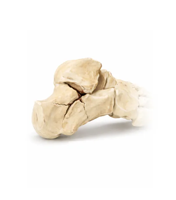
Calcaneal fractures are characterized by changes not only in bone integrity but also in the shape of the hindfoot, which directly influences gait mechanics. The calcaneus plays a key role in transmitting load from the lower leg to the foot and in shock absorption during walking, so its injury is often associated with alterations in heel height and width. Even if the bone heals, failure to restore these parameters may result in pain during weight-bearing and difficulty with prolonged walking.
The condition of the subtalar joint is particularly important, as it enables the foot to adapt to uneven surfaces. When the articular surface is involved, the foot’s ability to distribute load evenly is compromised. We assess the fracture pattern and the degree of deformity to select a treatment strategy aimed at preserving shock absorption and foot stability during movement.
Changes in the calcaneal angle and potential overload of other parts of the foot are also taken into account. Therefore, in calcaneal fractures, it is essential not only to achieve bone union but also to restore the anatomical shape of the heel to maintain proper gait biomechanics.
Restoration of weight-bearing capacity and foot function after a calcaneal fracture
In a calcaneal fracture, the treatment outcome largely depends on how well the foot retains its ability to absorb load during walking. Even after bone union, changes in heel shape or limited mobility of the subtalar joint may lead to pain with weight-bearing, stiffness, and rapid fatigue during prolonged walking. For the patient, this often means discomfort during normal daily activities.
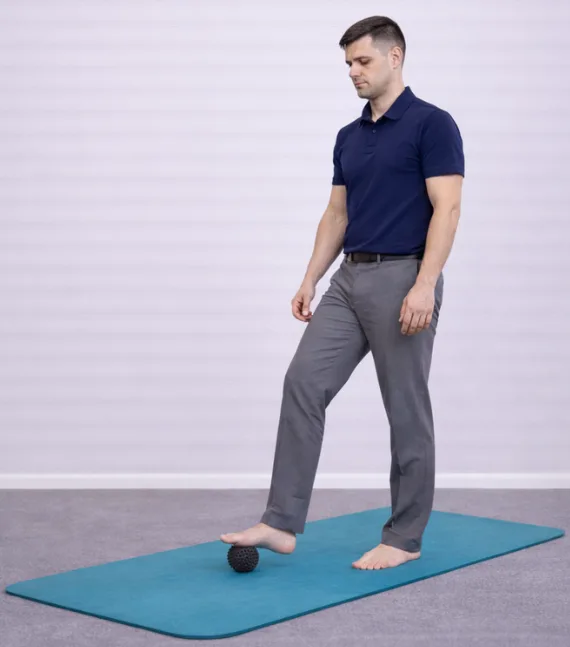
After osteosynthesis, it is important to gradually restore the foot’s shock-absorbing function and adapt it to load. We focus on regaining hindfoot mobility, ensuring even pressure distribution, and establishing a stable gait pattern. Premature overload or, conversely, prolonged protection may negatively affect the result, so recovery is structured in stages and carefully monitored.
We support the patient throughout the rehabilitation phase, assessing load tolerance and gait mechanics. This approach helps reduce the risk of chronic pain, maintain comfort while walking, and restore mobility without significant limitations.
Stages of treatment
Online consultation
An online consultation allows you to discuss your condition, complaints, and symptoms in advance. You can submit imaging studies and medical documents, ask questions, and receive a preliminary treatment plan before visiting the clinic.
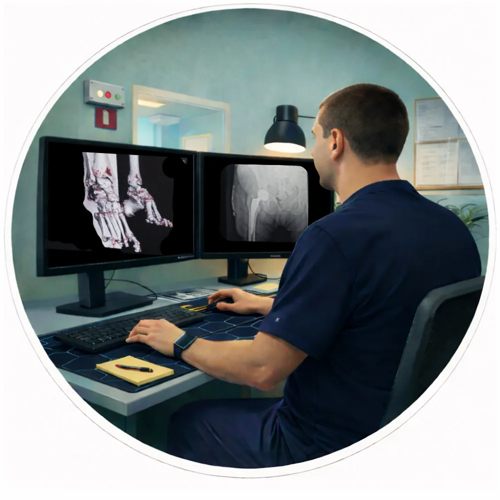
Examination and 3D surgical planning
All necessary examinations are performed within one day. A CT scan of the joint is conducted, followed by computer-based 3D surgical planning and implant selection to ensure maximum precision.

Surgery
On the second day of the clinic stay, the surgical procedure is performed. Original implants by Stryker Corporation (USA) and the MAKO robotic-assisted system are used, ensuring a high level of precision in implant positioning.
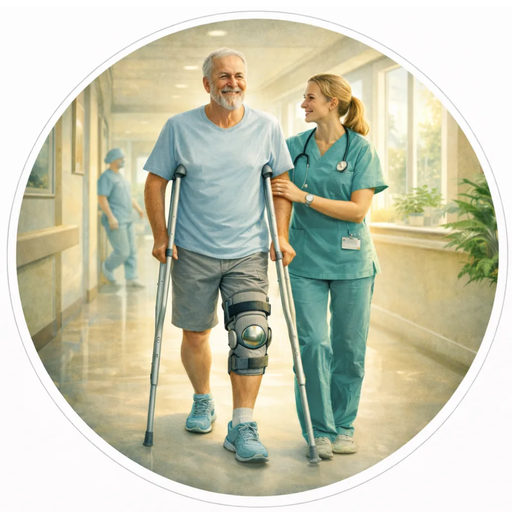
Rehabilitation and follow-up
Recovery begins immediately after surgery. In most cases, you will be able to start walking on the day of the procedure. After discharge, we continue to provide online follow-up care, including recovery guidance, rehabilitation monitoring, and answers to any questions that may arise.
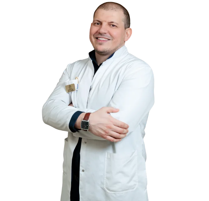
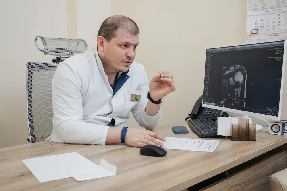
The surgery is performed personally by Zaur Sulumov
The doctor is personally involved in every stage of treatment — from the initial consultation and surgical planning to recovery monitoring and outcome evaluation.
Start Treatment
FAQ
The duration depends on the type of fracture and the fixation method. On average, surgery lasts from 30 to 120 minutes. In complex or comminuted fractures, operative time may be longer.
The timing of weight-bearing depends on the fracture location and stability of fixation. In some cases, movement is allowed within the first days after surgery, while full weight-bearing depends on the fracture type. For intra-articular fractures, a forced pause of 4–8 weeks may be required, with gradual progression under medical supervision and follow-up X-rays.
There is no pain during the operation due to anesthesia. After surgery, discomfort is managed with a modern pain-control protocol and typically decreases as the fracture heals.
Not always. Implants from leading manufacturers are commonly used, and in most cases plates and screws can remain in the body permanently. Removal is considered individually if there is discomfort or specific medical indications.
Average bone healing time ranges from 6 to 12 weeks, but this may vary depending on the patient’s age, fracture type, and overall health condition.
Rehabilitation focuses on restoring movement, muscle strength, and limb function. The program is individualized and may include physical therapy, physiotherapy, and gradual increase of load.
Not all fractures require surgical treatment. However, in cases of fragment displacement, instability, or risk of improper healing, osteosynthesis ensures accurate alignment and improves the functional outcome.